A critical part of the MNK and WND physiological response are their ability to change intracellular localisation in response to copper levels, relocating to sites where copper transport is required.
Under basal physiological copper levels, MNK and WND concentrate within the trans Golgi network (TGN) region, where they are postulated to pump copper into the TGN lumen for incorporatio into proteins on the secretory pathway.
When copper levels are raised, MNK has been reported to traffic to the plasma membrane and to the basolateral membrane in some polarized cell.
In response to elevated copper levels WND traffics to sub apical vesicles in some polarized cell lines, and has also been deonstrated partially at the apical membrane.
When intracellular copper levels are reduced, both transporters return via an endocytic route to the TGN.
Showing posts with label My project. Show all posts
Showing posts with label My project. Show all posts
Tuesday, 20 November 2007


My project (12)
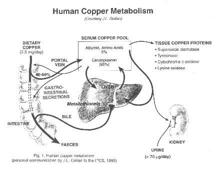
Human copper metabolism
Ingested copper is absorp from the gut 40-50%.
Copper bind to protein (mostly ceruloplasmin 95%).
Copper is transported to liver. Copper is secreted to bile and excreted to the gut again in feces
My project (11)
There are 13 types of ATP-ase
Na+/K+ transporting: ATP1A1, ATP1A2, ATP1A3, ATP1A4, ATP1B1, ATP1B2, ATP1B3, ATP1B4
Ca++ transporting: ATP2A1, ATP2A2, ATP2A3, ATP2B1, ATP2B2, ATP2B3, ATP2B4, ATP2C1
Mg++ transporting: ATP3
H+/K+ exchanging: ATP4A, ATP4B
H+ transporting, mitochondrial: ATP5A1, ATP5B, ATP5C1, ATP5C2, ATP5D, ATP5E, ATP5F1, ATP5G1, ATP5G2, ATP5G3, ATP5H, ATP5I, ATP5J, ATP5J2, ATP5L, ATP5L2, ATP5O, ATP5S
H+ transporting, lysosomal: ATP6AP1, ATP6AP2, ATP6V1A, ATP6V1B1, ATP6V1B2, ATP6V1C1, ATP6V1C2, ATP6V1D, ATP6V1E1, ATP6V1E2, ATP6V1F, ATP6V1G1, ATP6V1G2, ATP6V1G3, ATP6V1H, ATP6V0A1, ATP6V0A2, ATP6V0A4, ATP6V0B, ATP6V0C, ATP6V0D1, ATP6V0D2, ATP6V0E
Cu++ transporting: ATP7A (see also ATP7A), ATP7B (see also ATP7B)
Class I, type 8: ATP8A1, ATP8B1, ATP8B2, ATP8B3, ATP8B4
Class II, type 9: ATP9A, ATP9B
Class V, type 10: ATP10A, ATP10B, ATP10D
Class VI, type 11: ATP11A, ATP11B, ATP11C
H+/K+ transporting, nongastric: ATP12A
type 13: ATP13A1, ATP13A2, ATP13A3, ATP13A4, ATP13A5
Na+/K+ transporting: ATP1A1, ATP1A2, ATP1A3, ATP1A4, ATP1B1, ATP1B2, ATP1B3, ATP1B4
Ca++ transporting: ATP2A1, ATP2A2, ATP2A3, ATP2B1, ATP2B2, ATP2B3, ATP2B4, ATP2C1
Mg++ transporting: ATP3
H+/K+ exchanging: ATP4A, ATP4B
H+ transporting, mitochondrial: ATP5A1, ATP5B, ATP5C1, ATP5C2, ATP5D, ATP5E, ATP5F1, ATP5G1, ATP5G2, ATP5G3, ATP5H, ATP5I, ATP5J, ATP5J2, ATP5L, ATP5L2, ATP5O, ATP5S
H+ transporting, lysosomal: ATP6AP1, ATP6AP2, ATP6V1A, ATP6V1B1, ATP6V1B2, ATP6V1C1, ATP6V1C2, ATP6V1D, ATP6V1E1, ATP6V1E2, ATP6V1F, ATP6V1G1, ATP6V1G2, ATP6V1G3, ATP6V1H, ATP6V0A1, ATP6V0A2, ATP6V0A4, ATP6V0B, ATP6V0C, ATP6V0D1, ATP6V0D2, ATP6V0E
Cu++ transporting: ATP7A (see also ATP7A), ATP7B (see also ATP7B)
Class I, type 8: ATP8A1, ATP8B1, ATP8B2, ATP8B3, ATP8B4
Class II, type 9: ATP9A, ATP9B
Class V, type 10: ATP10A, ATP10B, ATP10D
Class VI, type 11: ATP11A, ATP11B, ATP11C
H+/K+ transporting, nongastric: ATP12A
type 13: ATP13A1, ATP13A2, ATP13A3, ATP13A4, ATP13A5


My project (8)
Intracellular levels of copper are in part controlled by two copper-transporting P-type ATPases, ATP7A(Menkes disease protein, MNK) and ATP7B(Wilson disease protein, WND). Both transporters are made up of 8 transmembrane domains and hydrolase ATP to translocate ions across cell membranes. MNK is involved in copper absorption, predominantly in the intestine and the kidney, whilst WND localizes predominantly to the liver and regulates copper clearanceby excreting it into bile.
MENKES DISEASE(#309400)
Clinical Synopsis
INHERITANCE :
X-linked recessive
GROWTH :
Height
Short stature
Other
Intrauterine growth retardation
HEAD AND NECK :
Head
Microcephaly
Brachycephaly
Wormian bones
Face
Pudgy cheeks
CARDIOVASCULAR :
Vascular
Intracranial hemorrhage
SKELETAL :
Osteoporosis
Skull
Wormian bones
Limbs
Metaphyseal widening with spurs
SKIN, NAILS, HAIR :
Skin
Hypopigmentation
Hair
Steely, kinky, sparse hair
Twisted and partial breaks on magnification
NEUROLOGIC :
Central nervous system
Degenerative neurologic disorder with onset age 1 month
Hypertonia
Seizures
Intracranial hemorrhage
Hypothermia
LABORATORY ABNORMALITIES :
Low copper and ceruloplasmin
WILSON DISEASE (#277900)
Clinical Synopsis
INHERITANCE :
Autosomal recessive
HEAD AND NECK :
Eyes
Kayser-Fleischer ring
ABDOMEN :
Liver
Atypical or prolonged hepatitis
Hepatic cirrhosis
Hepatic coma
Hepatomegaly
Liver failure
High liver copper
Gastrointestinal
Esophageal varices
GENITOURINARY :
Kidneys
Renal tubular dysfunction
Renal calculi
SKELETAL :
Osteoporosis
Osteomalacia
Chondrocalcinosis
Limbs
Osteoarthritis
Joint hypermobility
NEUROLOGIC :
Central nervous system
Tremor
Dysarthria
Dysphagia
Personality changes
Dementia
Poor motor coordination
Dystonia
Drooling
Peripheral nervous system
Mixed demyelinating and axonal polyneuropathy (rare)
ENDOCRINE FEATURES :
Hypoparathyroidism
HEMATOLOGY :
Hemolytic anemia
LABORATORY ABNORMALITIES :
Low serum ceruloplasmin
High nonceruloplasmin-bound serum copper
High urinary copper
Proteinuria
Aminoaciduria
Glycosuria
Uricaciduria
Hyperphosphaturia
Hypercalciuria
MISCELLANEOUS :
Incidence in United States of 1 in 55,000
Incidence worldwide of 1 in 30,000 to 50,000
MENKES DISEASE(#309400)
Clinical Synopsis
INHERITANCE :
X-linked recessive
GROWTH :
Height
Short stature
Other
Intrauterine growth retardation
HEAD AND NECK :
Head
Microcephaly
Brachycephaly
Wormian bones
Face
Pudgy cheeks
CARDIOVASCULAR :
Vascular
Intracranial hemorrhage
SKELETAL :
Osteoporosis
Skull
Wormian bones
Limbs
Metaphyseal widening with spurs
SKIN, NAILS, HAIR :
Skin
Hypopigmentation
Hair
Steely, kinky, sparse hair
Twisted and partial breaks on magnification
NEUROLOGIC :
Central nervous system
Degenerative neurologic disorder with onset age 1 month
Hypertonia
Seizures
Intracranial hemorrhage
Hypothermia
LABORATORY ABNORMALITIES :
Low copper and ceruloplasmin
WILSON DISEASE (#277900)
Clinical Synopsis
INHERITANCE :
Autosomal recessive
HEAD AND NECK :
Eyes
Kayser-Fleischer ring
ABDOMEN :
Liver
Atypical or prolonged hepatitis
Hepatic cirrhosis
Hepatic coma
Hepatomegaly
Liver failure
High liver copper
Gastrointestinal
Esophageal varices
GENITOURINARY :
Kidneys
Renal tubular dysfunction
Renal calculi
SKELETAL :
Osteoporosis
Osteomalacia
Chondrocalcinosis
Limbs
Osteoarthritis
Joint hypermobility
NEUROLOGIC :
Central nervous system
Tremor
Dysarthria
Dysphagia
Personality changes
Dementia
Poor motor coordination
Dystonia
Drooling
Peripheral nervous system
Mixed demyelinating and axonal polyneuropathy (rare)
ENDOCRINE FEATURES :
Hypoparathyroidism
HEMATOLOGY :
Hemolytic anemia
LABORATORY ABNORMALITIES :
Low serum ceruloplasmin
High nonceruloplasmin-bound serum copper
High urinary copper
Proteinuria
Aminoaciduria
Glycosuria
Uricaciduria
Hyperphosphaturia
Hypercalciuria
MISCELLANEOUS :
Incidence in United States of 1 in 55,000
Incidence worldwide of 1 in 30,000 to 50,000
Abstract for presentation at 11th International Congress of Human Genetics
Dr Objoon Trachoo, Division of Medical Genetics and Molecular Medicine, Department of Medicine, Ramathibodi Hospital, Mahidol University, Bangkok, Thailand
Dr Pimjai Nipharak, Division of Hematology, Department of Medicine, Ramathibodi Hospital, Mahidol University, Bangkok, Thailand
Mrs Kanoknan Srichan, Division of Medical Genetics and Molecular Medicine, Department of Medicine, Ramathibodi Hospital, Mahidol University, Bangkok, Thailand
Dr Prasit Phowthongkum, Division of Medical Genetics, Department of Medicine, King Chulalongkorn Memorial Hospital, Chulalongkorn University, Bangkok, Thailand
Dr Suporn Chanjarunee, Division of Hematology, Department of Medicine, Ramathibodi Hospital, Mahidol University, Bangkok, Thailand
Dr Surasak Kantachuvessiri, Division of Nephrology, Department of Medicine, Ramathibodi Hospital, Mahidol University, Bangkok, Thailand
Dr Panus Chalermsaenyakorn, Department of Pathology, Department of Medicine, Ramathibodi Hospital, Mahidol University, Bangkok, Thailand
A/Prof Thanyachai Sura, Division of Medical Genetics and Molecular Medicine, Department of Medicine, Ramathibodi Hospital, Mahidol University, Bangkok, Thailand
We report on Thai male identical twins who presented with hypochromic microcytic anemia, hepatosplenomegaly and gross hematuria since the age of 16. Hematological and hemoglobin analysis were consistent with a clinical diagnosis of beta thalassemia-Hb E disease and molecular diagnosis was confirmed by direct gene sequencing of all exons and splice junctions of beta globin gene. Neither structural renal disease nor renal stone was found from ultrasonographical findings. However, no specific cause of hematuria has been detected; therefore, renal biopsy was performed. Histological examination and specific immunological stain of renal tissue showed the evidence of IgA nephropathy. Recently, there were few reports of thalassemia disease associated with such particular renal involvement; thus far, we propose to include urinalysis screening in beta thalassemia-Hb E patients during follow-up in order to prevent further renal damage and end-stage renal disease.
Dr Pimjai Nipharak, Division of Hematology, Department of Medicine, Ramathibodi Hospital, Mahidol University, Bangkok, Thailand
Mrs Kanoknan Srichan, Division of Medical Genetics and Molecular Medicine, Department of Medicine, Ramathibodi Hospital, Mahidol University, Bangkok, Thailand
Dr Prasit Phowthongkum, Division of Medical Genetics, Department of Medicine, King Chulalongkorn Memorial Hospital, Chulalongkorn University, Bangkok, Thailand
Dr Suporn Chanjarunee, Division of Hematology, Department of Medicine, Ramathibodi Hospital, Mahidol University, Bangkok, Thailand
Dr Surasak Kantachuvessiri, Division of Nephrology, Department of Medicine, Ramathibodi Hospital, Mahidol University, Bangkok, Thailand
Dr Panus Chalermsaenyakorn, Department of Pathology, Department of Medicine, Ramathibodi Hospital, Mahidol University, Bangkok, Thailand
A/Prof Thanyachai Sura, Division of Medical Genetics and Molecular Medicine, Department of Medicine, Ramathibodi Hospital, Mahidol University, Bangkok, Thailand
We report on Thai male identical twins who presented with hypochromic microcytic anemia, hepatosplenomegaly and gross hematuria since the age of 16. Hematological and hemoglobin analysis were consistent with a clinical diagnosis of beta thalassemia-Hb E disease and molecular diagnosis was confirmed by direct gene sequencing of all exons and splice junctions of beta globin gene. Neither structural renal disease nor renal stone was found from ultrasonographical findings. However, no specific cause of hematuria has been detected; therefore, renal biopsy was performed. Histological examination and specific immunological stain of renal tissue showed the evidence of IgA nephropathy. Recently, there were few reports of thalassemia disease associated with such particular renal involvement; thus far, we propose to include urinalysis screening in beta thalassemia-Hb E patients during follow-up in order to prevent further renal damage and end-stage renal disease.
Monday, 19 November 2007
My project (7)
The homeostasis of copper within the body needs to be tightly regulated as deficiency prevents the proper functioning of proteins reliant on copper as a cofactor, whilst excess levels of copper are toxic cells.
Copper is an essential component of many metalloproteins, for example superoxide dismutase (free radical protection), mitochondrial cytochrome-c oxidase (electron transport), tyrosinase (pigmentation) and lysyl oxidase (collagen processing)
Copper is an essential component of many metalloproteins, for example superoxide dismutase (free radical protection), mitochondrial cytochrome-c oxidase (electron transport), tyrosinase (pigmentation) and lysyl oxidase (collagen processing)
Sunday, 18 November 2007
My project (6)
…to regulate trafflicking…
trafficking --> processing posttranslational modification of proteins, particularly secretory proteins and proteins targeted for membranes or specific cellular locations.
Source: http://www.biology-online.org/dictionary/Trafficking
trafficking --> processing posttranslational modification of proteins, particularly secretory proteins and proteins targeted for membranes or specific cellular locations.
Source: http://www.biology-online.org/dictionary/Trafficking
My project (5)
…ATP 7B…
*606882
ATPase, Cu(2+)-TRANSPORTING, BETA POLYPEPTIDE; ATP7B
Gene map locus 13q14.3-q21
DESCRIPTION
The ATP7B gene encodes a polypeptide that acts as a membrane copper-transport protein.
CLONING
On YACs from the 13q14.3 region, Bull et al. (1993) identified a sequence similar to that coding for the proposed copper-binding regions of the putative ATPase gene defective in Menkes disease (MNK; 309400). They showed that this sequence forms part of a P-type ATPase gene that is very similar to MNK, with 6 putative metal-binding regions similar to those found in prokaryotic heavy metal transporters. The gene, designated ATP7B, is expressed in liver and kidney, and was found to lie within a 300-kb region likely to include the Wilson disease locus (WND). The identity between MNK and the newly identified ATP7B gene was 78% in the transduction region, 89% in the channel/phosphorylation regions, and 79% in the ATP-binding region. The predicted length of the gene product was 1,411 amino acids for ATP7B compared with 1,500 amino acids for MNK. The overall identity between the two was 57%.
Tanzi et al. (1993) used consensus DNA sequences for heavy metal binding motifs to identify homologous cDNA clones. One of these was mapped by PCR amplification and Southern blotting to contiguous YAC and cosmid clones that span the WND locus at chromosome 13q14.3. The cDNA detected a 7.5-kb RNA transcript expressed most strongly in liver and brain. Sequence analysis indicated several membrane-spanning domains, an ATPase consensus sequence, a hinge domain, a phosphorylation site, and at least 2 putative copper-binding sites. The deduced protein showed 62% amino acid homology to the Menkes disease gene.
Yang et al. (1997) stated that the full-length ATP7B protein contains 1,465 amino acids. They cloned a splice variant of ATP7B lacking exons 6, 7, 8, and 12 from a human brain cDNA library. The deduced protein contains 1,258 amino acids. Immunofluorescence localization and fractionation of a hepatoma cell line revealed that the full-length protein was associated with the Golgi apparatus and the shorter isoform was cytosolic. Full-length ATP7B did not redistribute in response to elevated copper levels, and it did not associate with the plasma membrane.
Source: http://www.ncbi.nlm.nih.gov/entrez/dispomim.cgi?id=606882
*606882
ATPase, Cu(2+)-TRANSPORTING, BETA POLYPEPTIDE; ATP7B
Gene map locus 13q14.3-q21
DESCRIPTION
The ATP7B gene encodes a polypeptide that acts as a membrane copper-transport protein.
CLONING
On YACs from the 13q14.3 region, Bull et al. (1993) identified a sequence similar to that coding for the proposed copper-binding regions of the putative ATPase gene defective in Menkes disease (MNK; 309400). They showed that this sequence forms part of a P-type ATPase gene that is very similar to MNK, with 6 putative metal-binding regions similar to those found in prokaryotic heavy metal transporters. The gene, designated ATP7B, is expressed in liver and kidney, and was found to lie within a 300-kb region likely to include the Wilson disease locus (WND). The identity between MNK and the newly identified ATP7B gene was 78% in the transduction region, 89% in the channel/phosphorylation regions, and 79% in the ATP-binding region. The predicted length of the gene product was 1,411 amino acids for ATP7B compared with 1,500 amino acids for MNK. The overall identity between the two was 57%.
Tanzi et al. (1993) used consensus DNA sequences for heavy metal binding motifs to identify homologous cDNA clones. One of these was mapped by PCR amplification and Southern blotting to contiguous YAC and cosmid clones that span the WND locus at chromosome 13q14.3. The cDNA detected a 7.5-kb RNA transcript expressed most strongly in liver and brain. Sequence analysis indicated several membrane-spanning domains, an ATPase consensus sequence, a hinge domain, a phosphorylation site, and at least 2 putative copper-binding sites. The deduced protein showed 62% amino acid homology to the Menkes disease gene.
Yang et al. (1997) stated that the full-length ATP7B protein contains 1,465 amino acids. They cloned a splice variant of ATP7B lacking exons 6, 7, 8, and 12 from a human brain cDNA library. The deduced protein contains 1,258 amino acids. Immunofluorescence localization and fractionation of a hepatoma cell line revealed that the full-length protein was associated with the Golgi apparatus and the shorter isoform was cytosolic. Full-length ATP7B did not redistribute in response to elevated copper levels, and it did not associate with the plasma membrane.
Source: http://www.ncbi.nlm.nih.gov/entrez/dispomim.cgi?id=606882
My project (4)
…ATP 7B…
*606882
ATPase, Cu(2+)-TRANSPORTING, BETA POLYPEPTIDE; ATP7B
Gene map locus 13q14.3-q21.1
DESCRIPTION
The ATP7B gene encodes a polypeptide that acts as a membrane copper-transport protein.
CLONING
On YACs from the 13q14.3 region, Bull et al. (1993) identified a sequence similar to that coding for the proposed copper-binding regions of the putative ATPase gene defective in Menkes disease (MNK; 309400). They showed that this sequence forms part of a P-type ATPase gene that is very similar to MNK, with 6 putative metal-binding regions similar to those found in prokaryotic heavy metal transporters. The gene, designated ATP7B, is expressed in liver and kidney, and was found to lie within a 300-kb region likely to include the Wilson disease locus (WND). The identity between MNK and the newly identified ATP7B gene was 78% in the transduction region, 89% in the channel/phosphorylation regions, and 79% in the ATP-binding region. The predicted length of the gene product was 1,411 amino acids for ATP7B compared with 1,500 amino acids for MNK. The overall identity between the two was 57%.
Tanzi et al. (1993) used consensus DNA sequences for heavy metal binding motifs to identify homologous cDNA clones. One of these was mapped by PCR amplification and Southern blotting to contiguous YAC and cosmid clones that span the WND locus at chromosome 13q14.3. The cDNA detected a 7.5-kb RNA transcript expressed most strongly in liver and brain. Sequence analysis indicated several membrane-spanning domains, an ATPase consensus sequence, a hinge domain, a phosphorylation site, and at least 2 putative copper-binding sites. The deduced protein showed 62% amino acid homology to the Menkes disease gene.
Yang et al. (1997) stated that the full-length ATP7B protein contains 1,465 amino acids. They cloned a splice variant of ATP7B lacking exons 6, 7, 8, and 12 from a human brain cDNA library. The deduced protein contains 1,258 amino acids. Immunofluorescence localization and fractionation of a hepatoma cell line revealed that the full-length protein was associated with the Golgi apparatus and the shorter isoform was cytosolic. Full-length ATP7B did not redistribute in response to elevated copper levels, and it did not associate with the plasma membrane.
Source: http://www.ncbi.nlm.nih.gov/entrez/dispomim.cgi?id=606882
*606882
ATPase, Cu(2+)-TRANSPORTING, BETA POLYPEPTIDE; ATP7B
Gene map locus 13q14.3-q21.1
DESCRIPTION
The ATP7B gene encodes a polypeptide that acts as a membrane copper-transport protein.
CLONING
On YACs from the 13q14.3 region, Bull et al. (1993) identified a sequence similar to that coding for the proposed copper-binding regions of the putative ATPase gene defective in Menkes disease (MNK; 309400). They showed that this sequence forms part of a P-type ATPase gene that is very similar to MNK, with 6 putative metal-binding regions similar to those found in prokaryotic heavy metal transporters. The gene, designated ATP7B, is expressed in liver and kidney, and was found to lie within a 300-kb region likely to include the Wilson disease locus (WND). The identity between MNK and the newly identified ATP7B gene was 78% in the transduction region, 89% in the channel/phosphorylation regions, and 79% in the ATP-binding region. The predicted length of the gene product was 1,411 amino acids for ATP7B compared with 1,500 amino acids for MNK. The overall identity between the two was 57%.
Tanzi et al. (1993) used consensus DNA sequences for heavy metal binding motifs to identify homologous cDNA clones. One of these was mapped by PCR amplification and Southern blotting to contiguous YAC and cosmid clones that span the WND locus at chromosome 13q14.3. The cDNA detected a 7.5-kb RNA transcript expressed most strongly in liver and brain. Sequence analysis indicated several membrane-spanning domains, an ATPase consensus sequence, a hinge domain, a phosphorylation site, and at least 2 putative copper-binding sites. The deduced protein showed 62% amino acid homology to the Menkes disease gene.
Yang et al. (1997) stated that the full-length ATP7B protein contains 1,465 amino acids. They cloned a splice variant of ATP7B lacking exons 6, 7, 8, and 12 from a human brain cDNA library. The deduced protein contains 1,258 amino acids. Immunofluorescence localization and fractionation of a hepatoma cell line revealed that the full-length protein was associated with the Golgi apparatus and the shorter isoform was cytosolic. Full-length ATP7B did not redistribute in response to elevated copper levels, and it did not associate with the plasma membrane.
Source: http://www.ncbi.nlm.nih.gov/entrez/dispomim.cgi?id=606882
My project (3)
…ATP 7A and…
*300011
ATPase, Cu(2+)-TRANSPORTING, ALPHA POLYPEPTIDE; ATP7A
Gene map locus Xq12-q13
The ATP7A gene was cloned as a candidate for the site of mutations causing Menkes disease (MNK; 309400) by 3 independent groups (Vulpe et al., 1993; Chelly et al., 1993; Mercer et al., 1993). By a database search of the predicted sequence, Vulpe et al. (1993) found strong homology to P-type ATPases, a family of integral membrane proteins that use an aspartylphosphate intermediate to transport cations across membranes. The protein was found to have the characteristics of a copper-binding protein. Northern blot analysis showed that the mRNA of the gene, which was symbolized 'MNK' before its precise nature was known, is present in a variety of cell types and tissues, except liver, in which expression is reduced or absent. The findings were consistent with the clinical observation that the liver is largely unaffected in Menkes disease and fails to accumulate excess copper.
Levinson et al. (1994) and Mercer et al. (1994) isolated the mouse homolog of the Menkes disease gene. The mouse protein shows 89% identity to the human protein, and both proteins contain 8 transmembrane domains.
GENE STRUCTURE
Tumer et al. (1995) determined that the ATP7A gene spans about 150 kb of genomic DNA and contains 23 exons. The ATG start codon is in the second exon. The ATP7A and ATP7B (606882) genes showed strikingly similar exonic structures, with almost identical structures starting from the fifth metal-binding domain, suggesting the presence of a common ancestor encoding 1, and possibly 2, metal-binding domains in addition to the ATPase 'core.'
Dierick et al. (1995) showed that the ATP7A gene contains 23 exons distributed over approximately 140 kb of genomic DNA. The authors showed that exon 10 is alternatively spliced. They found that the structures of the ATP7A and ATP7B genes are similar in the 3-prime two-thirds region, consistent with their common evolutionary ancestry.
Source: http://www.ncbi.nlm.nih.gov/entrez/dispomim.cgi?id=300011
*300011
ATPase, Cu(2+)-TRANSPORTING, ALPHA POLYPEPTIDE; ATP7A
Gene map locus Xq12-q13
The ATP7A gene was cloned as a candidate for the site of mutations causing Menkes disease (MNK; 309400) by 3 independent groups (Vulpe et al., 1993; Chelly et al., 1993; Mercer et al., 1993). By a database search of the predicted sequence, Vulpe et al. (1993) found strong homology to P-type ATPases, a family of integral membrane proteins that use an aspartylphosphate intermediate to transport cations across membranes. The protein was found to have the characteristics of a copper-binding protein. Northern blot analysis showed that the mRNA of the gene, which was symbolized 'MNK' before its precise nature was known, is present in a variety of cell types and tissues, except liver, in which expression is reduced or absent. The findings were consistent with the clinical observation that the liver is largely unaffected in Menkes disease and fails to accumulate excess copper.
Levinson et al. (1994) and Mercer et al. (1994) isolated the mouse homolog of the Menkes disease gene. The mouse protein shows 89% identity to the human protein, and both proteins contain 8 transmembrane domains.
GENE STRUCTURE
Tumer et al. (1995) determined that the ATP7A gene spans about 150 kb of genomic DNA and contains 23 exons. The ATG start codon is in the second exon. The ATP7A and ATP7B (606882) genes showed strikingly similar exonic structures, with almost identical structures starting from the fifth metal-binding domain, suggesting the presence of a common ancestor encoding 1, and possibly 2, metal-binding domains in addition to the ATPase 'core.'
Dierick et al. (1995) showed that the ATP7A gene contains 23 exons distributed over approximately 140 kb of genomic DNA. The authors showed that exon 10 is alternatively spliced. They found that the structures of the ATP7A and ATP7B genes are similar in the 3-prime two-thirds region, consistent with their common evolutionary ancestry.
Source: http://www.ncbi.nlm.nih.gov/entrez/dispomim.cgi?id=300011
My project (2)

….proteins that interact with…
Protein-protein interactions refer to the association of protein molecules and the study of these associations from the perspective of biochemistry, signal transduction and networks.
Protein-protein interactions refer to the association of protein molecules and the study of these associations from the perspective of biochemistry, signal transduction and networks.
My project (1)
Identification of proteins….
Basics of protein identification are simple: Proteins are digested and the resulting peptides are analyzed by mass spectrometers to obtain their mass data. There are three different types of MS data that can be used for database search. They are
(1) molecular weights of peptides that can be used for Peptide Mass Mapping,
(2) combination of mass data and partial amino acid sequence that can be used for Sequence Tag, and
(3) tandem mass spectrometry data (uninterpreted) that are used for MS/MS fragmentation ion search.
Source: http://msf.ucdavis.edu/ms_protein_id.html
Basics of protein identification are simple: Proteins are digested and the resulting peptides are analyzed by mass spectrometers to obtain their mass data. There are three different types of MS data that can be used for database search. They are
(1) molecular weights of peptides that can be used for Peptide Mass Mapping,
(2) combination of mass data and partial amino acid sequence that can be used for Sequence Tag, and
(3) tandem mass spectrometry data (uninterpreted) that are used for MS/MS fragmentation ion search.
Source: http://msf.ucdavis.edu/ms_protein_id.html
Subscribe to:
Posts (Atom)