ความผิดปกติของฟันแบบพันธุกรรมไม่เข้ากลุ่มอาการ non syndromic inheritable teeth abnormalities
1. Dentinogenesis imperfecta (DI)
ถ่ายทอดแบบ AD
ชุดฟันที่เป็นปัญหา ทั้งฟันน้ำนมและฟันแท้
ลักษณะฟัน เทา เหลือง น้ำตาล ตัวฟันกว้าง คอแคบ เหมือนดอกทิวลิป อีนาเมล เปราะ แตกง่าย เหลือแต่เนื้อฟัน (Dentin)
ลักษณะรังสี ฟันเป็นของแข็งทึบ ไม่มีโพรง ไม่มีช่องรากฟัน
ลักษณะอื่นที่พบ บางรายมีหูหนวก (DFNA39 mutation ร่วมด้วย)
ตำแหน่งยีน โครโมโซม คู่ที่ 4 4q21.3 ยีน code dentin sialophosphoprotein (DSPP) ซึ่งเป็นโปรตีนที่เป็นองค์ประกอบสำคัญมากกว่า 50 % ในเนื้อฟัน ในส่วนที่ไม่ใช่คอลลาเจน A15V, P17T, V18F, Q45X
Differential diagnosis กับ osteogenesis imperfecta แต่โรคนี้มีแต่ผิดปกติที่ฟัน
การจำแนกชนิด
1. DI type I with OI (Osteogenesis imperfecta)
2. DI type II without OI
3. DI type III พบน้อยมาก และลักษณะกลับกันคือ เนื้อฟันมากไปเหมือนเปลือกหอย แต่ก็พบการกลายพันธุ์บนยีนที่ตำแหน่งเดียวกันกับ type II (allelic variant)
2. Dentin Dysplasia type I and II (DD I and DD II)
Type I ชื่ออื่น Rootless teeth, Radicular Dentin Dysplasia
ถ่ายทอดแบบ AD, มีรายงาน AR homozygous mutation ของยีน NFIC
ตำแหน่งยีน ยังไม่ทราบ
ลักษณะฟัน ภายนอก จะมีสีและรูปร่างค่อนข้างปกติ อาจจะสีเงาๆ ออกฟ้า หรือ น้ำตาลเล็กน้อยได้บ้าง
อาจเป็นฝีได้
ลักษณะ systemic มีรายงานพบลักษณะฟันแบบนี้ในคนไข้ที่มีลักษณะของ EDS type III (hypermobility syndrome ได้) มีรายงาน sclerotic ของ long bone ได้
ลักษณะทางรังสี ไม่มีรากฟันเลยและ มีรอยดำรอบๆ apical area หรือมีรากสั้นๆ ไปจนกระทั่งมีรากยาวปกติ แต่โพรงฟันแคบมาก และอาจมีหินปูนในโพรงฟัน (pulp)
Type II OMIM #125420 (Gene DSPP 125485)
ชื่ออื่น
DTDP2DENTIN DYSPLASIA, SHIELDS TYPE IICORONAL DENTIN DYSPLASIAANOMALOUS DYSPLASIA OF DENTINPULPAL DYSPLASIAPULP STONES
ถ่ายทอดแบบ AD น่าจะเป็น allelic variant ของ DI-II พบในตระกูลเดียวกันได้
ตำแหน่งยีน 4q21.3 DSPP gene Y6D (Rajpar et. al 2002)
ลักษณะฟัน ฟันน้ำนมจะมีสีเหลือง โปร่งแสง และไม่มีโพรงฟันเลย ส่วนฟันแท้จะมีสีภายนอกปกติ รากปกติ แต่โพรงฟันขึ้นไปถึง ตัวฟันด้านบน บานออกคล้ายเปลวไฟ หรือ thistle tube
Showing posts with label genetic disorders. Show all posts
Showing posts with label genetic disorders. Show all posts
Thursday, 6 March 2008


Thursday, 22 November 2007
Treament of genetic diseases in the real world 9: familial hypokalemic periodic paralysis
What is hypokalemic periodic paralysis?
Hypokalemic periodic paralysis is a condition that causes episodes of extreme muscle weakness typically beginning in childhood or adolescence. Most often, these episodes involve a temporary inability to move muscles in the arms and legs. Attacks cause severe weakness or paralysis that usually lasts from hours to days. Some people may have episodes almost every day, while others experience them weekly, monthly, or only rarely. Attacks can occur without warning or can be triggered by factors such as rest after exercise, a viral illness, or certain medications. Often, a large, carbohydrate-rich meal or vigorous exercise in the evening can trigger an attack upon waking the following morning. Although affected individuals usually regain their muscle strength between attacks, repeated episodes can lead to persistent muscle weakness later in life.
People with hypokalemic periodic paralysis have reduced levels of potassium in their blood (hypokalemia) during episodes of muscle weakness. Researchers are investigating how low potassium levels may be related to the muscle abnormalities in this condition.
How common is hypokalemic periodic paralysis?
Although its exact prevalence is unknown, hypokalemic periodic paralysis is estimated to affect 1 in 100,000 people. Men tend to experience symptoms of this condition more often than women.
Diagnosis/testing. The diagnosis of HOKPP is based on a history of episodes of flaccid paralysis; low serum concentration of potassium (<0.9 to 3.0 mmol/L) during attacks; the absence of myotonia clinically and on electromyography (EMG) (with the exception of one family with heat-induced myotonia and cold-induced HOKPP); and a family history consistent with autosomal dominant inheritance. Molecular genetic testing identifies disease-causing mutations in CACNA1S or SCN4A in 80% of individuals meeting clinical diagnostic criteria. Of all individuals with HOKPP, about 55-70% have mutations in CACNA1S and about 8-10% in SCN4A. Such testing is clinically available.
Management. Treatment of a paralytic crisis by administration of potassium by mouth or IV aims to normalize the serum concentration of potassium and to shorten the paralytic episode. ECG and blood potassium concentration must be monitored during treatment. Surveillance depends on the affected individual's symptoms and response to preventive treatment. Neurologic examination should focus on muscle strength in the legs to detect permanent weakness associated with myopathy. A number of factors can trigger paralytic attacks: unusually strenuous effort, excess of carbohydrate-rich meals, sweets, and alcohol should be avoided; oral or intravenous corticosteroids should be used with care; glucose infusion should be replaced by another type of infusion.
Potassium in doses of 0.2 to 0.4 mmol/kg is administered orally every 15 to 30 minutes over one to three hours.
If the individual is unable to swallow or does not tolerate potassium by mouth, potassium may be administered intravenously. In that case, it must be diluted in 5% mannitol rather than in glucose or sodium chloride, which trigger crises in individuals with HOKPP. The concentration of potassium administered intravenously must not exceed 40 mmol/L and the flow must not exceed 20 mmol/hour or 200-250 mmol/day; administration must be stopped when the serum potassium concentration is normalized, even if weakness persists.
Because the hypokalemia and subsequent changes in potassium
Genetic counseling. HOKPP is inherited in an autosomal dominant manner. Most individuals diagnosed with HOKPP have an affected parent. The proportion of cases caused by a de novo gene mutation is unknown. Offspring of a proband have a 50% risk of inheriting the mutation. Penetrance is about 90% in males and may be as low as 50% in females depending on the causative mutation. Prenatal testing is possible if the disease-causing mutation has been identified in the proband; however, requests for prenatal testing for conditions such as HOKPP that do not affect intellect and have some treatment available are not common.
POTASSIUM
Hypokalemic periodic paralysis is a condition that causes episodes of extreme muscle weakness typically beginning in childhood or adolescence. Most often, these episodes involve a temporary inability to move muscles in the arms and legs. Attacks cause severe weakness or paralysis that usually lasts from hours to days. Some people may have episodes almost every day, while others experience them weekly, monthly, or only rarely. Attacks can occur without warning or can be triggered by factors such as rest after exercise, a viral illness, or certain medications. Often, a large, carbohydrate-rich meal or vigorous exercise in the evening can trigger an attack upon waking the following morning. Although affected individuals usually regain their muscle strength between attacks, repeated episodes can lead to persistent muscle weakness later in life.
People with hypokalemic periodic paralysis have reduced levels of potassium in their blood (hypokalemia) during episodes of muscle weakness. Researchers are investigating how low potassium levels may be related to the muscle abnormalities in this condition.
How common is hypokalemic periodic paralysis?
Although its exact prevalence is unknown, hypokalemic periodic paralysis is estimated to affect 1 in 100,000 people. Men tend to experience symptoms of this condition more often than women.
Diagnosis/testing. The diagnosis of HOKPP is based on a history of episodes of flaccid paralysis; low serum concentration of potassium (<0.9 to 3.0 mmol/L) during attacks; the absence of myotonia clinically and on electromyography (EMG) (with the exception of one family with heat-induced myotonia and cold-induced HOKPP); and a family history consistent with autosomal dominant inheritance. Molecular genetic testing identifies disease-causing mutations in CACNA1S or SCN4A in 80% of individuals meeting clinical diagnostic criteria. Of all individuals with HOKPP, about 55-70% have mutations in CACNA1S and about 8-10% in SCN4A. Such testing is clinically available.
Management. Treatment of a paralytic crisis by administration of potassium by mouth or IV aims to normalize the serum concentration of potassium and to shorten the paralytic episode. ECG and blood potassium concentration must be monitored during treatment. Surveillance depends on the affected individual's symptoms and response to preventive treatment. Neurologic examination should focus on muscle strength in the legs to detect permanent weakness associated with myopathy. A number of factors can trigger paralytic attacks: unusually strenuous effort, excess of carbohydrate-rich meals, sweets, and alcohol should be avoided; oral or intravenous corticosteroids should be used with care; glucose infusion should be replaced by another type of infusion.
Potassium in doses of 0.2 to 0.4 mmol/kg is administered orally every 15 to 30 minutes over one to three hours.
If the individual is unable to swallow or does not tolerate potassium by mouth, potassium may be administered intravenously. In that case, it must be diluted in 5% mannitol rather than in glucose or sodium chloride, which trigger crises in individuals with HOKPP. The concentration of potassium administered intravenously must not exceed 40 mmol/L and the flow must not exceed 20 mmol/hour or 200-250 mmol/day; administration must be stopped when the serum potassium concentration is normalized, even if weakness persists.
Because the hypokalemia and subsequent changes in potassium
Genetic counseling. HOKPP is inherited in an autosomal dominant manner. Most individuals diagnosed with HOKPP have an affected parent. The proportion of cases caused by a de novo gene mutation is unknown. Offspring of a proband have a 50% risk of inheriting the mutation. Penetrance is about 90% in males and may be as low as 50% in females depending on the causative mutation. Prenatal testing is possible if the disease-causing mutation has been identified in the proband; however, requests for prenatal testing for conditions such as HOKPP that do not affect intellect and have some treatment available are not common.
POTASSIUM
Treatment of genetic diseases in tha real world 8: Hemochromatosis
Disease characteristics. HFE-associated hereditary hemochromatosis (HFE-HHC) is characterized by inappropriately high absorption of iron by the gastrointestinal mucosa, resulting in excessive storage of iron particularly in the liver, skin, pancreas, heart, joints, and testes. Abdominal pain, weakness, lethargy, and weight loss are early symptoms. Without therapy, males may develop symptoms between age 40 and 60 years and females after menopause. Hepatic fibrosis or cirrhosis may occur in untreated individuals after age 40 years. Other findings in untreated individuals may include progressive increase in skin pigmentation, diabetes mellitus, congestive heart failure and/or arrhythmias, arthritis, and hypogonadism.--> -->This description applies to individuals with clinical expression of HFE-HHC. A large, but yet as undefined, fraction of homozygotes for HFE-HHC do not develop clinical symptoms (i.e., penetrance is low).
Diagnosis/testing. The diagnosis of HFE-HHC in individuals with clinical symptoms consistent with HFE-HHC and/or biochemical evidence of iron overload is typically based on the results of the screening tests transferrin-iron saturation and serum ferritin concentration, and of confirmatory tests such as molecular genetic testing for the p.C282Y and p.H63D mutations in the HFE gene and/or histologic assessment of hepatic iron stores on liver biopsy. A threshold transferrin-iron saturation of 45% may be more sensitive for detecting HFE-HHC than the higher values used in the past. Although serum ferritin concentration may increase progressively over time in untreated individuals with HFE-HHC, it is not specific for HFE-HHC and cannot be used alone for identification of individuals with HFE-HHC. About 87% of individuals of European origin with HFE-HHC are either homozygotes for the p.C282Y mutation or compound heterozygotes for the p.C282Y and p.H63D mutations.
Management. Evaluations at initial diagnosis: liver biopsy in individuals with serum ferritin concentration greater than 1000 ng/mL to determine if cirrhosis is present. Treatment of manifestations: There is no general agreement that phlebotomy (removal of blood) treatment is indicated in the presence of biochemically defined abnormalities (i.e., elevated transferrin-iron saturation and elevated serum ferritin concentration) and the absence of characteristic clinical endpoints (i.e., diabetes mellitus, cirrhosis, and liver carcinoma). Since the long-term clinical course appears benign in the majority of those who have abnormal laboratory tests only, phlebotomy may be deferred; biannual follow-up testing for increasingly abnormal serum ferritin concentration and transferrin-iron saturation levels is recommended. In the presence of characteristic clinical endpoints, treatment by phlebotomy is indicated to maintain serum ferritin concentration at 50 ng/mL or lower. If affected individuals are identified before hepatic cirrhosis develops and if total body iron depletion is successfully accomplished by therapeutic phlebotomy, life expectancy approaches normal.
Genetic counseling. HFE-HHC is inherited in an autosomal recessive manner. Usually the genetic risk to sibs of a proband of having HFE-HHC is 25%. However, the high carrier frequency for a mutant HFE allele in the general population of European origin (11% of the population, or 1/9 persons) means that on occasion one parent has two abnormal HFE alleles, usually in the absence of clinical findings. In such instances, the risk to each sib of a proband of being homozygous for HFE-HHC is 50%. Offspring of an individual with HFE-HHC inherit one mutant HFE allele from the affected parent. Because the chance that the other parent is a carrier for a mutant HFE allele is 1/9, the risk to the offspring of having HFE-HHC is about 5%. Although prenatal testing would be technically feasible when both parents carry identified HFE mutations, such requests would be highly unusual because HFE-HHC is an adult-onset, treatable disease and the homozygous p.C282Y mutation has low clinical penetrance.
THERAPEUTIC PHLEBOTOMY
Diagnosis/testing. The diagnosis of HFE-HHC in individuals with clinical symptoms consistent with HFE-HHC and/or biochemical evidence of iron overload is typically based on the results of the screening tests transferrin-iron saturation and serum ferritin concentration, and of confirmatory tests such as molecular genetic testing for the p.C282Y and p.H63D mutations in the HFE gene and/or histologic assessment of hepatic iron stores on liver biopsy. A threshold transferrin-iron saturation of 45% may be more sensitive for detecting HFE-HHC than the higher values used in the past. Although serum ferritin concentration may increase progressively over time in untreated individuals with HFE-HHC, it is not specific for HFE-HHC and cannot be used alone for identification of individuals with HFE-HHC. About 87% of individuals of European origin with HFE-HHC are either homozygotes for the p.C282Y mutation or compound heterozygotes for the p.C282Y and p.H63D mutations.
Management. Evaluations at initial diagnosis: liver biopsy in individuals with serum ferritin concentration greater than 1000 ng/mL to determine if cirrhosis is present. Treatment of manifestations: There is no general agreement that phlebotomy (removal of blood) treatment is indicated in the presence of biochemically defined abnormalities (i.e., elevated transferrin-iron saturation and elevated serum ferritin concentration) and the absence of characteristic clinical endpoints (i.e., diabetes mellitus, cirrhosis, and liver carcinoma). Since the long-term clinical course appears benign in the majority of those who have abnormal laboratory tests only, phlebotomy may be deferred; biannual follow-up testing for increasingly abnormal serum ferritin concentration and transferrin-iron saturation levels is recommended. In the presence of characteristic clinical endpoints, treatment by phlebotomy is indicated to maintain serum ferritin concentration at 50 ng/mL or lower. If affected individuals are identified before hepatic cirrhosis develops and if total body iron depletion is successfully accomplished by therapeutic phlebotomy, life expectancy approaches normal.
Genetic counseling. HFE-HHC is inherited in an autosomal recessive manner. Usually the genetic risk to sibs of a proband of having HFE-HHC is 25%. However, the high carrier frequency for a mutant HFE allele in the general population of European origin (11% of the population, or 1/9 persons) means that on occasion one parent has two abnormal HFE alleles, usually in the absence of clinical findings. In such instances, the risk to each sib of a proband of being homozygous for HFE-HHC is 50%. Offspring of an individual with HFE-HHC inherit one mutant HFE allele from the affected parent. Because the chance that the other parent is a carrier for a mutant HFE allele is 1/9, the risk to the offspring of having HFE-HHC is about 5%. Although prenatal testing would be technically feasible when both parents carry identified HFE mutations, such requests would be highly unusual because HFE-HHC is an adult-onset, treatable disease and the homozygous p.C282Y mutation has low clinical penetrance.
THERAPEUTIC PHLEBOTOMY
Treament of genetic diseases in a real world 7: Wilson disease
See clinical synopsis in this link: wilson disease
Wilson disease can be treated effectively with metal chelating agent:
d-penicillamine (Cuprimine) especially in mild or asymptomatic cases, but patients with neurological symptoms way be worsen in the early period after treatment, and trientine(Syprine )is recommended for acute neurological alterations. Long term side effects of d-penicillamine is nephrotic syndrome, skin disruption and immune and bone marrow suppression. Zinc suphate is used in asymptomatic case or in maintenance phase after d-penicillamine treatment.
Agents/Circumstances to Avoid
Foods very high in copper (liver, brain, chocolate, mushrooms, shellfish, nuts), especially at the beginning of treatment
Wilson disease can be treated effectively with metal chelating agent:
d-penicillamine (Cuprimine) especially in mild or asymptomatic cases, but patients with neurological symptoms way be worsen in the early period after treatment, and trientine(Syprine )is recommended for acute neurological alterations. Long term side effects of d-penicillamine is nephrotic syndrome, skin disruption and immune and bone marrow suppression. Zinc suphate is used in asymptomatic case or in maintenance phase after d-penicillamine treatment.
Agents/Circumstances to Avoid
Foods very high in copper (liver, brain, chocolate, mushrooms, shellfish, nuts), especially at the beginning of treatment
Wednesday, 21 November 2007
Treatment of genetic diseases: in the real world. 5. Marfan syndrome
Marfan Syndrome is a multisystem inherited diseases; the major organ involvement is cardiovascular system. patient s with Marfan Syndrome will gradually develop enlargment of the great artery of heart (Aorta) due to weakness of the supporting tissues and leading to tearing (dissection) which are mostly fatal. See more informations of Marfan Syndrome in the link provided
http://clinicalgenetics.blogspot.com/2007/11/marfan-syndrome.html
Treatment of Marfan Syndrome is including advice to avoid vigorous exercise and regularly taking drug which reduce the force of heart beat that are proved to slow progression of the disease. Echocardiography (Ultrasound image of the heart and arteries) periodically to measure sizes of arteries in case of too much enlarge that hhaving high risk for tearing or rupture, prophylactic surgery will have done.
Drug use in Marfan Syndrome
Propanolol Inderal from Wyeth NEngl J Med. 1994 May 12;330(19):1335-41
Enalapril Renitec from Merck Am J Cardiol 2005;95:1125–1127
Atenolo and losartan
A Clinical Trial Comparing Atenolol and Losartan Is Beginning
Reed E. Pyeritz, M.D., Ph.D.
Professor of Medicine & Genetics
University of Pennsylvania School of Medicine
Philadelphia, PA, USA
http://clinicalgenetics.blogspot.com/2007/11/marfan-syndrome.html
Treatment of Marfan Syndrome is including advice to avoid vigorous exercise and regularly taking drug which reduce the force of heart beat that are proved to slow progression of the disease. Echocardiography (Ultrasound image of the heart and arteries) periodically to measure sizes of arteries in case of too much enlarge that hhaving high risk for tearing or rupture, prophylactic surgery will have done.
Drug use in Marfan Syndrome
Propanolol Inderal from Wyeth NEngl J Med. 1994 May 12;330(19):1335-41
Enalapril Renitec from Merck Am J Cardiol 2005;95:1125–1127
Atenolo and losartan
A Clinical Trial Comparing Atenolol and Losartan Is Beginning
Reed E. Pyeritz, M.D., Ph.D.
Professor of Medicine & Genetics
University of Pennsylvania School of Medicine
Philadelphia, PA, USA
Treatment of genetic diseases: in the real world.4. klinefelter's syndrome
What is Klinefelter syndrome?
Klinefelter syndrome is a chromosomal condition that affects male sexual development. Most males with Klinefelter syndrome have one extra copy of the X chromosome in each cell. Because their testicles do not develop normally, affected males may have low levels of the hormone testosterone beginning during puberty. A lack of this hormone can lead to breast development (gynecomastia), reduced facial and body hair, and an inability to father children (infertility). Compared with other men, adult males with Klinefelter syndrome have an increased risk of developing breast cancer and a chronic inflammatory disease called systemic lupus erythematosus. Their chance of developing these disorders is similar to that of normal adult females.
Boys with Klinefelter syndrome may have learning disabilities and difficulty with speech and language development. They tend to be quiet, sensitive, and unassertive, but personality characteristics vary among males with this condition.
Variants of Klinefelter syndrome, which involve more than one extra X chromosome or extra copies of both the X and Y chromosomes in each cell, tend to have more severe signs and symptoms. These disorders affect male sexual development and are associated with decreased IQ, distinctive facial features, skeletal abnormalities, poor coordination, and severe problems with speech.
How common is Klinefelter syndrome?
Klinefelter syndrome affects 1 in 500 to 1,000 males. Variants of Klinefelter syndrome are much rarer, occurring in 1 in 50,000 or fewer male births. Females are not affected by Klinefelter syndrome
Initial treatment
Treatment for Klinefelter syndrome usually starts when a boy is about 11 to 12 years old. It begins with measuring the amount of testosterone and other hormones in his blood. Many teenage boys with Klinefelter syndrome don't have low levels of testosterone.4 If a boy's testosterone level is low, he is given a man-made form of testosterone (Depo-Testosterone) on a regular basis. Testosterone can be given as an injection or through a skin patch or gel.
Testosterone:
Increases body hair, mainly on the face (beard), under the arm (axillary), and in the genital area (pubic).
Increases muscle development.
Increases sex drive.
Helps prevent osteoporosis.
May prevent or shrink enlarged breasts.
Provides better self-esteem by allowing the boy to "fit in" with his peers. This can result in more successful interpersonal relationships.
Side effects of testosterone replacement therapy are uncommon, but include worsening acne, overly rapid sexual development, and behavior problems (such as being overly aggressive). Careful monitoring is important to prevent these side effects.
Ongoing treatment
Ongoing treatment forKlinefelter syndrome may include:
Help for language and learning difficulties. If language delays are identified in early childhood, educational assistance and speech therapy can be used to treat the problem. Children with learning difficulties can receive educational support through the school system.
Testosterone replacement. This is given by injection or through a skin patch or gel. Testosterone replacement usually continues throughout the man's life but does not help infertility. For men who want to start a family, counseling and treatment at a fertility clinic are generally recommended.
Regular medical checkups to monitor for development of other conditions, such as autoimmune diseases, behavior problems, or psychiatric disorders, such as depression. Professional counseling or medication may be needed.
DEPO-TESTOSTERONE from Pfizer
Klinefelter syndrome is a chromosomal condition that affects male sexual development. Most males with Klinefelter syndrome have one extra copy of the X chromosome in each cell. Because their testicles do not develop normally, affected males may have low levels of the hormone testosterone beginning during puberty. A lack of this hormone can lead to breast development (gynecomastia), reduced facial and body hair, and an inability to father children (infertility). Compared with other men, adult males with Klinefelter syndrome have an increased risk of developing breast cancer and a chronic inflammatory disease called systemic lupus erythematosus. Their chance of developing these disorders is similar to that of normal adult females.
Boys with Klinefelter syndrome may have learning disabilities and difficulty with speech and language development. They tend to be quiet, sensitive, and unassertive, but personality characteristics vary among males with this condition.
Variants of Klinefelter syndrome, which involve more than one extra X chromosome or extra copies of both the X and Y chromosomes in each cell, tend to have more severe signs and symptoms. These disorders affect male sexual development and are associated with decreased IQ, distinctive facial features, skeletal abnormalities, poor coordination, and severe problems with speech.
How common is Klinefelter syndrome?
Klinefelter syndrome affects 1 in 500 to 1,000 males. Variants of Klinefelter syndrome are much rarer, occurring in 1 in 50,000 or fewer male births. Females are not affected by Klinefelter syndrome
Initial treatment
Treatment for Klinefelter syndrome usually starts when a boy is about 11 to 12 years old. It begins with measuring the amount of testosterone and other hormones in his blood. Many teenage boys with Klinefelter syndrome don't have low levels of testosterone.4 If a boy's testosterone level is low, he is given a man-made form of testosterone (Depo-Testosterone) on a regular basis. Testosterone can be given as an injection or through a skin patch or gel.
Testosterone:
Increases body hair, mainly on the face (beard), under the arm (axillary), and in the genital area (pubic).
Increases muscle development.
Increases sex drive.
Helps prevent osteoporosis.
May prevent or shrink enlarged breasts.
Provides better self-esteem by allowing the boy to "fit in" with his peers. This can result in more successful interpersonal relationships.
Side effects of testosterone replacement therapy are uncommon, but include worsening acne, overly rapid sexual development, and behavior problems (such as being overly aggressive). Careful monitoring is important to prevent these side effects.
Ongoing treatment
Ongoing treatment forKlinefelter syndrome may include:
Help for language and learning difficulties. If language delays are identified in early childhood, educational assistance and speech therapy can be used to treat the problem. Children with learning difficulties can receive educational support through the school system.
Testosterone replacement. This is given by injection or through a skin patch or gel. Testosterone replacement usually continues throughout the man's life but does not help infertility. For men who want to start a family, counseling and treatment at a fertility clinic are generally recommended.
Regular medical checkups to monitor for development of other conditions, such as autoimmune diseases, behavior problems, or psychiatric disorders, such as depression. Professional counseling or medication may be needed.
DEPO-TESTOSTERONE from Pfizer
Treatment of genetic diseases: in the real world. 3. Turner syndrome
What is Turner syndrome?
Turner syndrome is a chromosomal condition that alters development in females. Women with this condition tend to be shorter than average and are usually unable to conceive a child (infertile) because of an absence of ovarian function. Other features of this condition that can vary among women who have Turner syndrome include: extra skin on the neck (webbed neck), puffiness or swelling (lymphedema) of the hands and feet, skeletal abnormalities, heart defects and kidney problems.
This condition occurs in about 1 in 2,500 female births worldwide, but is much more common among pregnancies that do not survive to term (miscarriages and stillbirths).
Turner syndrome is a chromosomal condition related to the X chromosome. Researchers have not yet determined which genes on the X chromosome are responsible for most signs and symptoms of Turner syndrome. They have, however, identified one gene called SHOX that is important for bone development and growth. Missing one copy of this gene likely causes short stature and skeletal abnormalities in women with Turner syndrome.
What is the treatment for Turner syndrome?
During childhood and adolescence, girls may be under the care of a pediatric endocrinologist, who is a specialist in childhood conditions of the hormones and metabolism.
Growth hormone injections are beneficial in some individuals with Turner syndrome. Injections often begin in early childhood and may increase final adult height by a few inches.
Estrogen replacement therapy is usually started at the time of normal puberty, around 12 years to start breast development. Estrogen and progesterone are given a little later to begin a monthly 'period,' which is necessary to keep the womb healthy. Estrogen is also given to prevent osteoporosis.
Babies born with a heart murmur or narrowing of the aorta may need surgery to correct the problem. A heart expert (cardiologist) will assess and follow up any treatment necessary.
Girls who have Turner syndrome are more likely to get middle ear infections. Repeated infections may lead to hearing loss and should be evaluated by the pediatrician. An ear, nose and throat specialist (ENT) may be involved in caring for this health issue.
High blood pressure is quite common in women who have Turner syndrome. In some cases, the elevated blood pressure is due to narrowing of the aorta or a kidney abnormality. However, most of the time, no specific cause for the elevation is identified. Blood pressure should be checked routinely and, if necessary, treated with medication. Women who have Turner syndrome have a slightly higher risk of having an under active thyroid or developing diabetes. This should also be monitored during routine health maintenance visits and treated if necessary.
Regular health checks are very important. Special clinics for the care of girls and women who have Turner syndrome are available in some areas, with access to a variety of specialists. Early preventive care and treatment is very important.
Almost all women are infertile, but pregnancy with donor embryos may be possible.
Drug used for Turner syndrome patients
Estrogen to increase height, produce secondary sexual charactristics, prevent osteoporois, improve fertility in some cases.
Premarin conjugated estrogen from Wyeth
Growth hormone to increase height.
Humatrope recombinant Human Growth Hormone from EliLily
Thyroid hormone in patients with hypothyroids
Eltroxin T4 Tetrathyroiodine from Glaxo Welcome
Turner syndrome is a chromosomal condition that alters development in females. Women with this condition tend to be shorter than average and are usually unable to conceive a child (infertile) because of an absence of ovarian function. Other features of this condition that can vary among women who have Turner syndrome include: extra skin on the neck (webbed neck), puffiness or swelling (lymphedema) of the hands and feet, skeletal abnormalities, heart defects and kidney problems.
This condition occurs in about 1 in 2,500 female births worldwide, but is much more common among pregnancies that do not survive to term (miscarriages and stillbirths).
Turner syndrome is a chromosomal condition related to the X chromosome. Researchers have not yet determined which genes on the X chromosome are responsible for most signs and symptoms of Turner syndrome. They have, however, identified one gene called SHOX that is important for bone development and growth. Missing one copy of this gene likely causes short stature and skeletal abnormalities in women with Turner syndrome.
What is the treatment for Turner syndrome?
During childhood and adolescence, girls may be under the care of a pediatric endocrinologist, who is a specialist in childhood conditions of the hormones and metabolism.
Growth hormone injections are beneficial in some individuals with Turner syndrome. Injections often begin in early childhood and may increase final adult height by a few inches.
Estrogen replacement therapy is usually started at the time of normal puberty, around 12 years to start breast development. Estrogen and progesterone are given a little later to begin a monthly 'period,' which is necessary to keep the womb healthy. Estrogen is also given to prevent osteoporosis.
Babies born with a heart murmur or narrowing of the aorta may need surgery to correct the problem. A heart expert (cardiologist) will assess and follow up any treatment necessary.
Girls who have Turner syndrome are more likely to get middle ear infections. Repeated infections may lead to hearing loss and should be evaluated by the pediatrician. An ear, nose and throat specialist (ENT) may be involved in caring for this health issue.
High blood pressure is quite common in women who have Turner syndrome. In some cases, the elevated blood pressure is due to narrowing of the aorta or a kidney abnormality. However, most of the time, no specific cause for the elevation is identified. Blood pressure should be checked routinely and, if necessary, treated with medication. Women who have Turner syndrome have a slightly higher risk of having an under active thyroid or developing diabetes. This should also be monitored during routine health maintenance visits and treated if necessary.
Regular health checks are very important. Special clinics for the care of girls and women who have Turner syndrome are available in some areas, with access to a variety of specialists. Early preventive care and treatment is very important.
Almost all women are infertile, but pregnancy with donor embryos may be possible.
Drug used for Turner syndrome patients
Estrogen to increase height, produce secondary sexual charactristics, prevent osteoporois, improve fertility in some cases.
Premarin conjugated estrogen from Wyeth
Growth hormone to increase height.
Humatrope recombinant Human Growth Hormone from EliLily
Thyroid hormone in patients with hypothyroids
Eltroxin T4 Tetrathyroiodine from Glaxo Welcome
Treatment of genetic diseases: in the real world. 2. Hemophilia
Hemophilia is an inherited bleeding disorders that was record since the ancient time.
The male new born has to be circumscribed for religious purpose, the affected individuals can be bleed until dead leading to exception for families with history of bleeding in male.
The most famous family with hemophilia is the Queen victorian royal family; there are many male suffered from this kind of severe bleeding disorders.
Once upon a time, those affected individulas died in before 10 years olf from sever intracranila bleeding. Nowadays, we know that these patients are lacking of clotting factor in their blood. Two common types of disease lack from differnet factor: factor 8 in hemophilia A and factor 9 in hemophilia B. These factors are found in normal individual plasma and that can be collected, concentrate and given to patiens in need. from the advance of genetic engineering, we can synthesized factor 8 or 9 with recombinant technology to produce large amount of factors independently for blood donors anymore, but they are still expensive and restricted for use in the most severe one.
These are the list of saving life products for hemophiliac patients:
Fresh Frozen Plasma
Cryoprecipitated (factor 8 rich)
Cryoremoved plasma (factor 9 rich)
Factor 7 recombinant: Novoseven from Novo Nordisk (can be used in both types)
Factor 8 recombinant: Advate from baxter, Helixate from Behring
Factor 9 recombinant: BeneFIX from Wyeth
Anti-Inhibitor Coagulant Complex FEIBA from Baxter (can be used in both types)
In mild hemophilia A or hemophiliac patients undegoing to perform oral procedure: transamin (tranexamic acid) from Daiichi
The male new born has to be circumscribed for religious purpose, the affected individuals can be bleed until dead leading to exception for families with history of bleeding in male.
The most famous family with hemophilia is the Queen victorian royal family; there are many male suffered from this kind of severe bleeding disorders.
Once upon a time, those affected individulas died in before 10 years olf from sever intracranila bleeding. Nowadays, we know that these patients are lacking of clotting factor in their blood. Two common types of disease lack from differnet factor: factor 8 in hemophilia A and factor 9 in hemophilia B. These factors are found in normal individual plasma and that can be collected, concentrate and given to patiens in need. from the advance of genetic engineering, we can synthesized factor 8 or 9 with recombinant technology to produce large amount of factors independently for blood donors anymore, but they are still expensive and restricted for use in the most severe one.
These are the list of saving life products for hemophiliac patients:
Fresh Frozen Plasma
Cryoprecipitated (factor 8 rich)
Cryoremoved plasma (factor 9 rich)
Factor 8 concentrate plasma
Factor 9 concentrate plasma
Factor 7 recombinant: Novoseven from Novo Nordisk (can be used in both types)
Factor 8 recombinant: Advate from baxter, Helixate from Behring
Factor 9 recombinant: BeneFIX from Wyeth
Anti-Inhibitor Coagulant Complex FEIBA from Baxter (can be used in both types)
In mild hemophilia A or hemophiliac patients undegoing to perform oral procedure: transamin (tranexamic acid) from Daiichi
Treatment of genetic diseases: in the real world. 1. familial hypercholesterolemia
One of the most common misunderstanding or negative attitudes towards genetic diseases is Genetic diseases can not be treated. They may sometimes hear about gene therapy, cloning or other advanced knowledge requirement to understand, so they leave genetics at the corner of their mind. But what do genetics doctors say?
I have to say that there are thousands of genetic diseases (and this long list is growing rapidly) that are treated or can be prevented with drugs, diet therapy, or surgical procedures effectively.
For emphasizing this promising areas of medicine, I will list these genetic diseases and their management that change their hopeless life to the bright new one.
The first one I'd like to mention is familial hypercholesterolemia.
Familial hypercholesterolemia
Familial hypercholesterolemia is one of the most common genetic diseases in the world; the prevalence of heterozygous (get one abnormal gene from only one parent) affected people is about 1 in 500, which equal to prevalence of stroke in England.
Affected people have consistenly high levels of low density lipoprotein (LDL or bad cholesterol) which leads to premature atherosclerosis of the coronary arteries, cerebral arteries and peripheral vessels. Typically in affected men, heart attacks occur in their 40s to 50s, and 85% of men with this disorder have experienced a heart attack by age 60. The incidence of heart attacks in women with this disorder is also increased, but happens 10 years later than in men.
In homozygous (receive abnormal genes from both parents) individuals the condition is more severe, and cholesterol values may exceed 600 mg/dl (normal not exceed 200 mg/dl) These individuals develop waxy plaques (xanthoma) beneath the skin over their elbows, knees, buttocks. These are deposits of cholesterol in the skin. In addition, they develop deposits in tendons, and around the cornea of the eye. atherosclerosis begins before pubery and heart attacks and death may occur before age30, or they may require higly invasive surgery such as a liver transplant. Fortunately homozygous individulas are extremely rare in about 1 in million.
Hetrozygous patients may respond well to diet modifications combined with strong lipid lowering agents which have a class name statins.
Statins are the most common and one of the most prescribed drugs nowadays for treatment of high blood cholesterol. Most of high blood cholesterol individuals are partially inherited, but not in the case of familial hypercholesterolmeia which transfer to the next generation at rate about 50 %.
There are many guidelines for managing affected individulas.
Statins provides excellent control of inherited high cholestrol levels. The statins work by reducing the manufacture of cholestrol by cells. This stimulated LDL receptor gene expression. As a result, the receptors produced by the normal gene wil reduce cholesterol levels. the effect of statins can be enhanced by bile acid sequestants or cholesterol absorption inhibitors such as plant sterols or the recently introduced drug, ezetimibe.
Many patients with FH can achieve target cholesterol levels.
STATIN TREATMENT OF FH MALES IS ONE OF HE MOST COST_EFFECTIVE MEDICAL INTERVENTIONS AVAILABLE AND SEVERAL LINE OF EVIDENCE POINT TOWARDS MAJOR IMPROVEMENTS IN CARDIOVASCULAR EVENT RATES AND TOTAL MORTALITY(DEAD) OF FH PATIENTS.
These are the names of drugs can be used for Familail Hypercholesterolemia:
Lipitor from Pfizer : Atorvastatin
Crestor from Aventis: Rosuvastatin
high dose Zocor from Merck: simvastatin
Ezetrol from Merck: ezetimibe in combination with Zocor
I have to say that there are thousands of genetic diseases (and this long list is growing rapidly) that are treated or can be prevented with drugs, diet therapy, or surgical procedures effectively.
For emphasizing this promising areas of medicine, I will list these genetic diseases and their management that change their hopeless life to the bright new one.
The first one I'd like to mention is familial hypercholesterolemia.
Familial hypercholesterolemia
Familial hypercholesterolemia is one of the most common genetic diseases in the world; the prevalence of heterozygous (get one abnormal gene from only one parent) affected people is about 1 in 500, which equal to prevalence of stroke in England.
Affected people have consistenly high levels of low density lipoprotein (LDL or bad cholesterol) which leads to premature atherosclerosis of the coronary arteries, cerebral arteries and peripheral vessels. Typically in affected men, heart attacks occur in their 40s to 50s, and 85% of men with this disorder have experienced a heart attack by age 60. The incidence of heart attacks in women with this disorder is also increased, but happens 10 years later than in men.
In homozygous (receive abnormal genes from both parents) individuals the condition is more severe, and cholesterol values may exceed 600 mg/dl (normal not exceed 200 mg/dl) These individuals develop waxy plaques (xanthoma) beneath the skin over their elbows, knees, buttocks. These are deposits of cholesterol in the skin. In addition, they develop deposits in tendons, and around the cornea of the eye. atherosclerosis begins before pubery and heart attacks and death may occur before age30, or they may require higly invasive surgery such as a liver transplant. Fortunately homozygous individulas are extremely rare in about 1 in million.
Hetrozygous patients may respond well to diet modifications combined with strong lipid lowering agents which have a class name statins.
Statins are the most common and one of the most prescribed drugs nowadays for treatment of high blood cholesterol. Most of high blood cholesterol individuals are partially inherited, but not in the case of familial hypercholesterolmeia which transfer to the next generation at rate about 50 %.
There are many guidelines for managing affected individulas.
Statins provides excellent control of inherited high cholestrol levels. The statins work by reducing the manufacture of cholestrol by cells. This stimulated LDL receptor gene expression. As a result, the receptors produced by the normal gene wil reduce cholesterol levels. the effect of statins can be enhanced by bile acid sequestants or cholesterol absorption inhibitors such as plant sterols or the recently introduced drug, ezetimibe.
Many patients with FH can achieve target cholesterol levels.
STATIN TREATMENT OF FH MALES IS ONE OF HE MOST COST_EFFECTIVE MEDICAL INTERVENTIONS AVAILABLE AND SEVERAL LINE OF EVIDENCE POINT TOWARDS MAJOR IMPROVEMENTS IN CARDIOVASCULAR EVENT RATES AND TOTAL MORTALITY(DEAD) OF FH PATIENTS.
These are the names of drugs can be used for Familail Hypercholesterolemia:
Lipitor from Pfizer : Atorvastatin
Crestor from Aventis: Rosuvastatin
high dose Zocor from Merck: simvastatin
Ezetrol from Merck: ezetimibe in combination with Zocor
Tuesday, 20 November 2007
My project (8)
Intracellular levels of copper are in part controlled by two copper-transporting P-type ATPases, ATP7A(Menkes disease protein, MNK) and ATP7B(Wilson disease protein, WND). Both transporters are made up of 8 transmembrane domains and hydrolase ATP to translocate ions across cell membranes. MNK is involved in copper absorption, predominantly in the intestine and the kidney, whilst WND localizes predominantly to the liver and regulates copper clearanceby excreting it into bile.
MENKES DISEASE(#309400)
Clinical Synopsis
INHERITANCE :
X-linked recessive
GROWTH :
Height
Short stature
Other
Intrauterine growth retardation
HEAD AND NECK :
Head
Microcephaly
Brachycephaly
Wormian bones
Face
Pudgy cheeks
CARDIOVASCULAR :
Vascular
Intracranial hemorrhage
SKELETAL :
Osteoporosis
Skull
Wormian bones
Limbs
Metaphyseal widening with spurs
SKIN, NAILS, HAIR :
Skin
Hypopigmentation
Hair
Steely, kinky, sparse hair
Twisted and partial breaks on magnification
NEUROLOGIC :
Central nervous system
Degenerative neurologic disorder with onset age 1 month
Hypertonia
Seizures
Intracranial hemorrhage
Hypothermia
LABORATORY ABNORMALITIES :
Low copper and ceruloplasmin
WILSON DISEASE (#277900)
Clinical Synopsis
INHERITANCE :
Autosomal recessive
HEAD AND NECK :
Eyes
Kayser-Fleischer ring
ABDOMEN :
Liver
Atypical or prolonged hepatitis
Hepatic cirrhosis
Hepatic coma
Hepatomegaly
Liver failure
High liver copper
Gastrointestinal
Esophageal varices
GENITOURINARY :
Kidneys
Renal tubular dysfunction
Renal calculi
SKELETAL :
Osteoporosis
Osteomalacia
Chondrocalcinosis
Limbs
Osteoarthritis
Joint hypermobility
NEUROLOGIC :
Central nervous system
Tremor
Dysarthria
Dysphagia
Personality changes
Dementia
Poor motor coordination
Dystonia
Drooling
Peripheral nervous system
Mixed demyelinating and axonal polyneuropathy (rare)
ENDOCRINE FEATURES :
Hypoparathyroidism
HEMATOLOGY :
Hemolytic anemia
LABORATORY ABNORMALITIES :
Low serum ceruloplasmin
High nonceruloplasmin-bound serum copper
High urinary copper
Proteinuria
Aminoaciduria
Glycosuria
Uricaciduria
Hyperphosphaturia
Hypercalciuria
MISCELLANEOUS :
Incidence in United States of 1 in 55,000
Incidence worldwide of 1 in 30,000 to 50,000
MENKES DISEASE(#309400)
Clinical Synopsis
INHERITANCE :
X-linked recessive
GROWTH :
Height
Short stature
Other
Intrauterine growth retardation
HEAD AND NECK :
Head
Microcephaly
Brachycephaly
Wormian bones
Face
Pudgy cheeks
CARDIOVASCULAR :
Vascular
Intracranial hemorrhage
SKELETAL :
Osteoporosis
Skull
Wormian bones
Limbs
Metaphyseal widening with spurs
SKIN, NAILS, HAIR :
Skin
Hypopigmentation
Hair
Steely, kinky, sparse hair
Twisted and partial breaks on magnification
NEUROLOGIC :
Central nervous system
Degenerative neurologic disorder with onset age 1 month
Hypertonia
Seizures
Intracranial hemorrhage
Hypothermia
LABORATORY ABNORMALITIES :
Low copper and ceruloplasmin
WILSON DISEASE (#277900)
Clinical Synopsis
INHERITANCE :
Autosomal recessive
HEAD AND NECK :
Eyes
Kayser-Fleischer ring
ABDOMEN :
Liver
Atypical or prolonged hepatitis
Hepatic cirrhosis
Hepatic coma
Hepatomegaly
Liver failure
High liver copper
Gastrointestinal
Esophageal varices
GENITOURINARY :
Kidneys
Renal tubular dysfunction
Renal calculi
SKELETAL :
Osteoporosis
Osteomalacia
Chondrocalcinosis
Limbs
Osteoarthritis
Joint hypermobility
NEUROLOGIC :
Central nervous system
Tremor
Dysarthria
Dysphagia
Personality changes
Dementia
Poor motor coordination
Dystonia
Drooling
Peripheral nervous system
Mixed demyelinating and axonal polyneuropathy (rare)
ENDOCRINE FEATURES :
Hypoparathyroidism
HEMATOLOGY :
Hemolytic anemia
LABORATORY ABNORMALITIES :
Low serum ceruloplasmin
High nonceruloplasmin-bound serum copper
High urinary copper
Proteinuria
Aminoaciduria
Glycosuria
Uricaciduria
Hyperphosphaturia
Hypercalciuria
MISCELLANEOUS :
Incidence in United States of 1 in 55,000
Incidence worldwide of 1 in 30,000 to 50,000
Thalassemia is a genetic disorders that are characterized with chronic anemia. The disease is cause from the gene that control hemoglobin production. Hemoglobins are proteins that are the major components of red blood cell. Human carries mutation of thalassemia genes may be asymptomatic or have mild anmia, or severe until they died before birth.
In Thailand, a south east Asian country, there are more than 30 percent having one mutated hemoglobin genes. We call this group as carrier. They will be healthy and asymptomatic, but they can inherited their mutated gene to their offsprings. If their couples are also the same type carriers, their off springs have chance to have mutated genes both from thier parents that will produce diseases. The number of thalassemia patients in Thailad is about 600000.
And each year, couples at risk (both are carriers) give thalassemic baby for more than 10000 annually.
Ther are two common type of thalassemia in Thailand: alpha-thalassemia and beta-thalassemia.
In general, everyone are inherited the genetic materials half from their father and the rest from their mother. Thalassemia is a disease transfered in recessive fashion, which means that you must have mutated genes from both parents to have disease. Besides, the mutation must be on the same gene. For example, alpha-thalassemia gene mutation should pair with alpha-thalassemia gene mutation to produce alpha-thalassemia patients. Who have one mutated gene of beta-type and onother mutated gene of alpha-type are healthy.
Thalassemia is the result of decreasing production of hemoglobin leading to unbalanced between each type of hemoglobin. In normal, two chain of alpha hemoglobin will be paired with two chains of beta hemoglobin. In the non-balancing situation, the excess hemoglobin will be aggregated and cause changing of red cell shape and vulnerability. red cell will be survived less, be easily destroy. These all lead to chronic anemia. Organ that produce red blood cell i.e. bone marrow, liver, and spleen have to be expanded for increasing production rate, so we will see these patients have a big and tall skull, fragile bone, big liver, big spleen. In severe cases, this occured early in thier life, and inevitable cause of death in their toddler. Milder case will be survived to their teen with regular blood transfusion. The drawbcak of blood transfusion is the iron stored in the blood cell will be accumulated in patient's heart, liver, skin, pancreas, and so on lead to malfunction of that organs that are fatal if lefted untreated. Nowadays, in the most severe type that will be dead before birth, we have technology to test the fetus and termination before birth or have complication will be done if test positive. In severe birth infants, we can treated with bone marrow transplantation, but this is a very expensive methods and there is a high death rate from this procedure and not everyone can performed these due to unmatched tissues. Milder case will receive regular blood transfusion to prevent abnormal growth and bone change, and iron-chelator will be given to prevent long term complication for iron-excess. Prevention is more appropriate practice, and prenatal screening method is the one of most success genetic project in our country. In the future , next four or five generation, we will have less thalassemia patients.
In Thailand, a south east Asian country, there are more than 30 percent having one mutated hemoglobin genes. We call this group as carrier. They will be healthy and asymptomatic, but they can inherited their mutated gene to their offsprings. If their couples are also the same type carriers, their off springs have chance to have mutated genes both from thier parents that will produce diseases. The number of thalassemia patients in Thailad is about 600000.
And each year, couples at risk (both are carriers) give thalassemic baby for more than 10000 annually.
Ther are two common type of thalassemia in Thailand: alpha-thalassemia and beta-thalassemia.
In general, everyone are inherited the genetic materials half from their father and the rest from their mother. Thalassemia is a disease transfered in recessive fashion, which means that you must have mutated genes from both parents to have disease. Besides, the mutation must be on the same gene. For example, alpha-thalassemia gene mutation should pair with alpha-thalassemia gene mutation to produce alpha-thalassemia patients. Who have one mutated gene of beta-type and onother mutated gene of alpha-type are healthy.
Thalassemia is the result of decreasing production of hemoglobin leading to unbalanced between each type of hemoglobin. In normal, two chain of alpha hemoglobin will be paired with two chains of beta hemoglobin. In the non-balancing situation, the excess hemoglobin will be aggregated and cause changing of red cell shape and vulnerability. red cell will be survived less, be easily destroy. These all lead to chronic anemia. Organ that produce red blood cell i.e. bone marrow, liver, and spleen have to be expanded for increasing production rate, so we will see these patients have a big and tall skull, fragile bone, big liver, big spleen. In severe cases, this occured early in thier life, and inevitable cause of death in their toddler. Milder case will be survived to their teen with regular blood transfusion. The drawbcak of blood transfusion is the iron stored in the blood cell will be accumulated in patient's heart, liver, skin, pancreas, and so on lead to malfunction of that organs that are fatal if lefted untreated. Nowadays, in the most severe type that will be dead before birth, we have technology to test the fetus and termination before birth or have complication will be done if test positive. In severe birth infants, we can treated with bone marrow transplantation, but this is a very expensive methods and there is a high death rate from this procedure and not everyone can performed these due to unmatched tissues. Milder case will receive regular blood transfusion to prevent abnormal growth and bone change, and iron-chelator will be given to prevent long term complication for iron-excess. Prevention is more appropriate practice, and prenatal screening method is the one of most success genetic project in our country. In the future , next four or five generation, we will have less thalassemia patients.
Common situation in genetic counseling
Today, a medical student asked me about her relative was pregnant and she found that her baby had thalassemia from the prenatal screening. Her doctor gave her a chance to terminate the pregnancy. This student shown me the genotype: Hb Malay (Mutation at codon 19)/ Hb E. This is called compound heterozygotes, in contrary to double hetrozygotes that have mutation on different genes. To answer this question, we should know about the severity of phenotypes, effect to mother.Fortunately, thalassemia is one of the most common and the most throughly study genetic diseases, so we have some information to cope with that. But genotype-phenotype correlation is not an easy issue, there are many factors beyond the mutation itself to correctly predict that. Like these situations, Hb Maly/Hb E infants are not severe (thalassemia intermedia), but the characteristics are widely range from asymptomatic, need some blood transfusion or more frequent need of blood transfusion until required iron chelating therapy or splenectomy in mid-life. There is no specific curative treatment for this group, while beta thallassemia major is so severe and will be died in first two or three years of life, so high mortality-procedure dependent like bone marrow transplantation is accepted.
So the best recommendation for this situation is give all the informations needed to make a good decision for those counselee i.e. severity of baby, effect for pregnancy, treatment plan for baby, side effect of termination, choices, etc. And let the families making their own decision because they have to know what is the best way for their future life. Don't use our standard, believes, social issues to force or coercion them.
So the best recommendation for this situation is give all the informations needed to make a good decision for those counselee i.e. severity of baby, effect for pregnancy, treatment plan for baby, side effect of termination, choices, etc. And let the families making their own decision because they have to know what is the best way for their future life. Don't use our standard, believes, social issues to force or coercion them.
Monday, 19 November 2007
Let's share experience about genetic disorders
This is a story of a families suffered from hereditary non polyposis colon cancer that was left undiagnoses for a long period.
The index patient is a 68 yr old women. She came to thyroid clinic as a regular basis to reevaluate her neck mass that was diagnosis later as benign hyroid nodule for about 2 years. Last time, my resident asked her about her family, and this resident found that this is a very interesting family. She was diagnosed transitional cell carcinoma of left kidneys and was undergone success total nephrectomy for 20 years. She also had undergone hysterectmy (cut out the womb) that was diagnosed benign smooth muscle tumor. Her brother was suffered from small intestinal adenocardinoma in his 40 years. She had 5 offsprings, two males and three females. Her son was healthy, her daughters are diagnosed ovarian cancer and colon cancer each. Left only one daughter in her 40 normal.
So this family had four members suffered from cancers which can be group to fit for hereditary non polyposis colon cancer syndrome, which is a cancer predisposing syndrome. Member who carried the abnormal gene have about 70% risk to developed colon cancer and a substantial risks to develop other tumors such as kidneys, ovaries, small intestines like this family.
The family memebers were arranged to see me. I gave them a new information that they never heard before. Surveillance colonoscopy (only the index patient go for virtual CT colonoscopy), pelvic ultrasound, serum tumor marker screening are recommended for patients and who are at 50% risk to carrier the mutated gene.
Genetic testing of this syndrome is very exhaustive, due to different genes (up to 4 or more) can be the cause of the syndrome, very large gene and many other limited factors, so this genetic test are not adopted as routine practice in our country.
The family member said " We feel that we are lucky to know this information. For more than 20 years, we see our relatives diagnosed with cancer and we cannot do anything. Now we know that genetics play important part and we can surveillance as a regular basis to find it and to manage it promptly. We are happier that we were."
The index patient is a 68 yr old women. She came to thyroid clinic as a regular basis to reevaluate her neck mass that was diagnosis later as benign hyroid nodule for about 2 years. Last time, my resident asked her about her family, and this resident found that this is a very interesting family. She was diagnosed transitional cell carcinoma of left kidneys and was undergone success total nephrectomy for 20 years. She also had undergone hysterectmy (cut out the womb) that was diagnosed benign smooth muscle tumor. Her brother was suffered from small intestinal adenocardinoma in his 40 years. She had 5 offsprings, two males and three females. Her son was healthy, her daughters are diagnosed ovarian cancer and colon cancer each. Left only one daughter in her 40 normal.
So this family had four members suffered from cancers which can be group to fit for hereditary non polyposis colon cancer syndrome, which is a cancer predisposing syndrome. Member who carried the abnormal gene have about 70% risk to developed colon cancer and a substantial risks to develop other tumors such as kidneys, ovaries, small intestines like this family.
The family memebers were arranged to see me. I gave them a new information that they never heard before. Surveillance colonoscopy (only the index patient go for virtual CT colonoscopy), pelvic ultrasound, serum tumor marker screening are recommended for patients and who are at 50% risk to carrier the mutated gene.
Genetic testing of this syndrome is very exhaustive, due to different genes (up to 4 or more) can be the cause of the syndrome, very large gene and many other limited factors, so this genetic test are not adopted as routine practice in our country.
The family member said " We feel that we are lucky to know this information. For more than 20 years, we see our relatives diagnosed with cancer and we cannot do anything. Now we know that genetics play important part and we can surveillance as a regular basis to find it and to manage it promptly. We are happier that we were."
Pain in the back, blood in the urine
Have anyone heard about ADPKD (autosomal dominant polycystic kidney disease), one of the most common genetic disorders in the world?
It is the disease of the kidneys, the pea-shape pairs organs, weight about 10 ounces each, in the back of the body on each side of your backbone. They filter your blood and produce urine to remove waste that we produce every day. They also regulates your blood chemicals and acid-base status. The other functions of them is regulation of blood cell production response to body need, produce active vitamin D for helping in calcium absorption to strengthening your bones. If they are severe damage or nearly loss of their function from any causes, so called kidney failure, you will become weakness, loss of energy, shortness of breath, nausea/vomiting, swelling, and even leading to death if kidney replacement such as dialysis, or kidney transplantation is performed. But if they loss only part of their functions, you may not have any symptoms. The most common causes of kidney failure are diabetes and hypertension, which affect adult and elderly and immune-mediated kidney dieases and ADPKD are the more common causes of kidney failure in teenage or young adults.
ADPKD is a kidney diseases characteristics with multiple, various in sizes, growing in both kidneys. The diseases are inherited from parents in a dominant fashion, that means when only one affected father or mother can produced offsprings with ADPKD ( risk is 50%). The defected gene is in every cells of the child since their birth, but usually cysts are gradual develop in their tens or twenties. The patients may come to medical attention by many presentations such as back pain, blood in urine, hypertension, mass in your belly, infection of urinary tract or accidental finding on other health problem unrelated to the disease.
The cysts will grow until they compress normal kidney tissues and this will lead to kidney failure in their 40s-50s. The patients will died if dialysis, or kidney transplantation is not given in time.
The dianosis can be made by perform the imaging study of the kidneys such as ultraasound or CT in patietns suspected to be ADPKD (previous lists of symptoms and family members affected with the same problems) or in family members of ADPKD. Molecular testing can be performed in some families to diagnosis presymptomatic and precyst formation family members.
Treatment of pain, infection and hypertension may alleviate the symptoms and slow down the groth rate of cysts. Due to its serious complications and they are quite common as high as 1 in 1000 birth, this is one of the most active research area to find the drugs to stop progression of diseases.
And we are nearly at that times to treat genetic diseases with simple drugs.
See more information of ADPKD in PKD foundation websites
It is the disease of the kidneys, the pea-shape pairs organs, weight about 10 ounces each, in the back of the body on each side of your backbone. They filter your blood and produce urine to remove waste that we produce every day. They also regulates your blood chemicals and acid-base status. The other functions of them is regulation of blood cell production response to body need, produce active vitamin D for helping in calcium absorption to strengthening your bones. If they are severe damage or nearly loss of their function from any causes, so called kidney failure, you will become weakness, loss of energy, shortness of breath, nausea/vomiting, swelling, and even leading to death if kidney replacement such as dialysis, or kidney transplantation is performed. But if they loss only part of their functions, you may not have any symptoms. The most common causes of kidney failure are diabetes and hypertension, which affect adult and elderly and immune-mediated kidney dieases and ADPKD are the more common causes of kidney failure in teenage or young adults.
ADPKD is a kidney diseases characteristics with multiple, various in sizes, growing in both kidneys. The diseases are inherited from parents in a dominant fashion, that means when only one affected father or mother can produced offsprings with ADPKD ( risk is 50%). The defected gene is in every cells of the child since their birth, but usually cysts are gradual develop in their tens or twenties. The patients may come to medical attention by many presentations such as back pain, blood in urine, hypertension, mass in your belly, infection of urinary tract or accidental finding on other health problem unrelated to the disease.
The cysts will grow until they compress normal kidney tissues and this will lead to kidney failure in their 40s-50s. The patients will died if dialysis, or kidney transplantation is not given in time.
The dianosis can be made by perform the imaging study of the kidneys such as ultraasound or CT in patietns suspected to be ADPKD (previous lists of symptoms and family members affected with the same problems) or in family members of ADPKD. Molecular testing can be performed in some families to diagnosis presymptomatic and precyst formation family members.
Treatment of pain, infection and hypertension may alleviate the symptoms and slow down the groth rate of cysts. Due to its serious complications and they are quite common as high as 1 in 1000 birth, this is one of the most active research area to find the drugs to stop progression of diseases.
And we are nearly at that times to treat genetic diseases with simple drugs.
See more information of ADPKD in PKD foundation websites


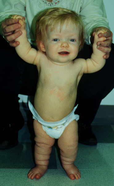

Achodroplasia
What is achondroplasia?
Achondroplasia is a disorder of bone growth. Although achondroplasia literally means "without cartilage formation," the problem is not in forming cartilage but in converting it to bone, particularly in the long bones of the arms and legs.
All people with achondroplasia have short stature. The average height of an adult male with achondroplasia is 131 centimeters (4 feet, 4 inches), and the average height for adult females is 124 centimeters (4 feet, 1 inch). Characteristic features of achondroplasia include an average-size trunk, short arms and legs with particularly short upper arms and thighs, limited range of motion at the elbows, and an enlarged head (macrocephaly) with a prominent forehead. Fingers are typically short and the ring finger and middle finger may diverge, giving the hand a three-pronged (trident) appearance. People with achondroplasia are generally of normal intelligence.
Health problems commonly associated with achondroplasia include episodes in which breathing slows or stops for short periods (apnea), obesity, and recurrent ear infections. In adulthood, individuals with the condition usually develop a pronounced and permanent sway of the lower back (lordosis) and bowed legs. Older individuals often have back pain, which can cause difficulty with walking.
How common is achondroplasia?
Achondroplasia is the most common type of short-limbed dwarfism. The condition occurs in 1 in 15,000 to 40,000 newborns.
What genes are related to achondroplasia?
Mutations in the FGFR3 gene cause achondroplasia.
The FGFR3 gene provides instructions for making a protein that is involved in the development and maintenance of bone and brain tissue. This protein limits the formation of bone from cartilage (a process called ossification), particularly in the long bones. Two specific mutations in the FGFR3 gene are responsible for almost all cases of achondroplasia. Researchers believe that these mutations cause the protein to be overly active, which interferes with skeletal development and leads to the disturbances in bone growth seen with this disorder.
How do people inherit achondroplasia?
Achondroplasia is inherited in an autosomal dominant pattern, which means one copy of the altered gene in each cell is sufficient to cause the disorder. About 80 percent of people with achondroplasia have average-size parents; these cases result from a new mutation in the FGFR3 gene. In the remaining cases, people with achondroplasia have inherited an altered FGFR3 gene from one or two affected parents. Individuals who inherit two altered copies of this gene typically have very severe problems with bone growth, and are usually stillborn or die shortly after birth from respiratory failure.
Achondroplasia is a disorder of bone growth. Although achondroplasia literally means "without cartilage formation," the problem is not in forming cartilage but in converting it to bone, particularly in the long bones of the arms and legs.
All people with achondroplasia have short stature. The average height of an adult male with achondroplasia is 131 centimeters (4 feet, 4 inches), and the average height for adult females is 124 centimeters (4 feet, 1 inch). Characteristic features of achondroplasia include an average-size trunk, short arms and legs with particularly short upper arms and thighs, limited range of motion at the elbows, and an enlarged head (macrocephaly) with a prominent forehead. Fingers are typically short and the ring finger and middle finger may diverge, giving the hand a three-pronged (trident) appearance. People with achondroplasia are generally of normal intelligence.
Health problems commonly associated with achondroplasia include episodes in which breathing slows or stops for short periods (apnea), obesity, and recurrent ear infections. In adulthood, individuals with the condition usually develop a pronounced and permanent sway of the lower back (lordosis) and bowed legs. Older individuals often have back pain, which can cause difficulty with walking.
How common is achondroplasia?
Achondroplasia is the most common type of short-limbed dwarfism. The condition occurs in 1 in 15,000 to 40,000 newborns.
What genes are related to achondroplasia?
Mutations in the FGFR3 gene cause achondroplasia.
The FGFR3 gene provides instructions for making a protein that is involved in the development and maintenance of bone and brain tissue. This protein limits the formation of bone from cartilage (a process called ossification), particularly in the long bones. Two specific mutations in the FGFR3 gene are responsible for almost all cases of achondroplasia. Researchers believe that these mutations cause the protein to be overly active, which interferes with skeletal development and leads to the disturbances in bone growth seen with this disorder.
How do people inherit achondroplasia?
Achondroplasia is inherited in an autosomal dominant pattern, which means one copy of the altered gene in each cell is sufficient to cause the disorder. About 80 percent of people with achondroplasia have average-size parents; these cases result from a new mutation in the FGFR3 gene. In the remaining cases, people with achondroplasia have inherited an altered FGFR3 gene from one or two affected parents. Individuals who inherit two altered copies of this gene typically have very severe problems with bone growth, and are usually stillborn or die shortly after birth from respiratory failure.
Down Syndrome
Introduction
Human cells normally contain 23 pairs of chromosomes. Chromosomes are the parts of body cells that carry inherited information - the characteristics that are passed to you from your mother and father.
A chromosomal disorder means there is a change in the normal number of chromosomes. This can mean that physical and mental development is impaired and can result in learning difficulties and health problems.
Downs Syndrome is the most common chromosomal disorder and one of the most common causes of learning difficulties.
Named after the first person that described it, Dr J L Down, the condition affects one in every 1,000 people. Some are more severely affected than others.
Around 600 babies with Downs Syndrome are born in the UK every year. It occurs in all ethnic groups and affects slightly more boys than girls.
Symptoms
People with Downs Syndrome have lots of different characteristics.
There are thought to be up to 120 features of Downs Syndrome but many children have no more than six or seven of them.
Physical features:
Babies with Downs Syndrome usually weigh less than average at birth and are usually shorter than average as adults.
Children with Downs Syndrome often have a rounded face, with a flat profile (the face looks flat when you view it from the side). The back of the head is slightly flattened (this is called brachycephaly) and the eyes tend to slant upwards.
There are small folds of skin that run vertically between the inner corner of the eye and the bridge of the nose (epicanthic folds) and this can give the impression of crossed eyes (squint). There may be white or yellow speckling around the rim of the iris (coloured part of the eye). These are called Brushfield spots.
Children with Downs Syndrome often have straight, soft hair. As children they may have an extra fold of skin over the back of the neck and as adults, short broad necks.
People with Downs Syndrome often have smaller than average mouths with a bigger than average tongue that may stick out.
The hands may be broad with short fingers; the little finger may only have one joint instead of two and be slightly curved. The feet are often stubby with a wide space between the first and second toes.
Many people with Downs Syndrome have poor muscle tone (hypotonia). This muscular floppiness occurs in the limbs and neck and usually improves with age.
Children with Downs Syndrome learn new skills more slowly than other children and generally develop at a slower rate, meeting their developmental milestones such as walking or talking later. They may progress in stops and starts and may not fully catch up with other children their age. Often, a child with Downs Syndrome will not start to use language until his or her third year and may use some sign language before talking.
With treatment and support, the average life expectancy of someone with Downs Syndrome is about 60 years.
Human cells normally contain 23 pairs of chromosomes. Chromosomes are the parts of body cells that carry inherited information - the characteristics that are passed to you from your mother and father.
A chromosomal disorder means there is a change in the normal number of chromosomes. This can mean that physical and mental development is impaired and can result in learning difficulties and health problems.
Downs Syndrome is the most common chromosomal disorder and one of the most common causes of learning difficulties.
Named after the first person that described it, Dr J L Down, the condition affects one in every 1,000 people. Some are more severely affected than others.
Around 600 babies with Downs Syndrome are born in the UK every year. It occurs in all ethnic groups and affects slightly more boys than girls.
Symptoms
People with Downs Syndrome have lots of different characteristics.
There are thought to be up to 120 features of Downs Syndrome but many children have no more than six or seven of them.
Physical features:
Babies with Downs Syndrome usually weigh less than average at birth and are usually shorter than average as adults.
Children with Downs Syndrome often have a rounded face, with a flat profile (the face looks flat when you view it from the side). The back of the head is slightly flattened (this is called brachycephaly) and the eyes tend to slant upwards.
There are small folds of skin that run vertically between the inner corner of the eye and the bridge of the nose (epicanthic folds) and this can give the impression of crossed eyes (squint). There may be white or yellow speckling around the rim of the iris (coloured part of the eye). These are called Brushfield spots.
Children with Downs Syndrome often have straight, soft hair. As children they may have an extra fold of skin over the back of the neck and as adults, short broad necks.
People with Downs Syndrome often have smaller than average mouths with a bigger than average tongue that may stick out.
The hands may be broad with short fingers; the little finger may only have one joint instead of two and be slightly curved. The feet are often stubby with a wide space between the first and second toes.
Many people with Downs Syndrome have poor muscle tone (hypotonia). This muscular floppiness occurs in the limbs and neck and usually improves with age.
Children with Downs Syndrome learn new skills more slowly than other children and generally develop at a slower rate, meeting their developmental milestones such as walking or talking later. They may progress in stops and starts and may not fully catch up with other children their age. Often, a child with Downs Syndrome will not start to use language until his or her third year and may use some sign language before talking.
With treatment and support, the average life expectancy of someone with Downs Syndrome is about 60 years.
Subscribe to:
Posts (Atom)